Archive for the ‘Dr. Raymond Stein’ Category

Meet the New Professor of Ophthalmology & Vision Sciences at U of T

Monday, May 24th, 2021
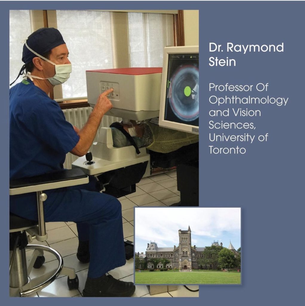
Dr. Raymond Stein, Professor of Ophthalmology and Vision Sciences at the University of Toronto

We are thrilled to announce that Dr. Raymond Stein, was promoted to Professor of Ophthalmology and Vision Sciences at the University of Toronto. This is the highest designation at the University in recognition of having an international impact and scholarly activities. Dr. Stein’s accomplishments include writing scientific articles, books, chapters, as well as presentations and seminars to students and other doctors. Over 30 professors of ophthalmology from around the world wrote letters of support for this promotion. Congratulations on this remarkable achievement Dr. Raymond Stein, of Bochner Eye Institute.

This Year’s Top Choice Winner for Laser Vision Correction

Thursday, March 4th, 2021

Top Choice in Toronto for LASIKDr. Raymond Stein and his team are pleased to announce that Bochner Eye Institute is Toronto’s 2021 Top Choice Winner for Laser Vision Correction. The recognition honors “consistent excellence in the pursuit of customer satisfaction.”

The award is based on surveys of hundreds of customers and members of the community. Their experiences are used to quantify “the trust and loyalty [businesses] have earned from the people they serve.” Winners display the proprietary Mark of Excellence and are listed in the iTrustLocal.com directory.

Dr. Stein is gratified that clients prize his depth of experience, the convenience and value of the laser vision correction procedures he offers, the extraordinary precision of his LASIK surgical technique and also the satisfying customer experience. This includes the friendliness of the staff, the transparency and integrity of the Institute’s core values and the personalized attention every patient receives.

Trust a Top Surgeon

As a trusted Toronto ophthalmologist, Dr. Stein’s commitment to our city and its communities runs deep. A graduate of the University of Toronto Medical School, Dr. Stein also served as Chief Ophthalmologist at the Scarborough Hospital and is currently Associate Professor of Ophthalmology at the University of Toronto. Although his reputation transcends the community, he always puts local patients first, providing advanced care and the most sophisticated surgical procedures to the families of our city.

A former president of the Canadian Society of Cataract and Refractive Surgery, Dr. Stein is a prolific author, popular lecturer and recognized innovator. He is also the current editor of Clinical and Surgical Ophthalmology, a highly respected medical journal. His years of training include an ophthalmology residency at the Mayo Clinic and a cornea and external diseases fellowship at the Wills Eye Hospital in Philadelphia.

He estimates he has performed nearly a quarter million surgical procedures to date, including cataract surgery, refractive lens exchange, LASIK and PRK. He is equally adept at routine vision correction procedures and complex cases involving keratoconus and loss of eyesight.

To schedule an appointment with experienced Toronto ophthalmologist Dr. Raymond Stein, please call or email Bochner Eye Institute today.

Dr. Raymond Stein Is Keynote Speaker at Ophthalmology Meeting at Columbia University in NYC

Saturday, January 12th, 2019

Dr. Raymond Stein was pleased to be a keynote speaker at the 2018 Precision Ophthalmology “Optimizing the Refractive Status of the Eye” meeting at Columbia University in New York City. Held November 30, the meeting gathered ophthalmologists, optometrists and eye care professionals from around North America to discuss the current state of cataract surgery, refractive surgery, dry eye and corneal disease treatments and share information about advances in the field.

In his first talk, Dr. Stein reflected on some of the lessons he has learned in 10 years of performing topography-guided PRK and corneal crosslinking for the treatment of keratoconus. Dr. Stein and his colleagues at Bochner Eye Institute are some of the first doctors in Canada to use this approach to treat the progressive thinning and bulging of the cornea. To date Dr. Stein has performed over 5,000 of these procedures with excellent results. 

Later, Dr. Stein spoke about using a corneal laser to treat age-related macular degeneration (AMD), or the deterioration of the portion of the retina responsible for central vision. Bochner Eye Institute is the first practice in North America to use a laser to modify the cornea and improve distance and near vision in patients with AMD. A study conducted by the Bochner team showed that patients who had the laser procedure experienced improvements in both distance and near vision three years after treatment.

Dr. Stein was invited to speak at the Precision Ophthalmology meeting by Dr. Stephen Trokel, a renowned ophthalmologist and a pioneer in refractive surgery. Dr. Trokel developed the excimer laser for vision correction and performed the first human excimer laser surgery in 1987. He also trained Dr. Stein in refractive surgery in 1991.

Earlier in 2018, Dr. Stein spoke at the World Ophthalmology Congress of the International Council of Ophthalmology in Barcelona as well as the Canadian Ophthalmological Society’s annual meeting, which took place in Toronto.

Contact Dr. Stein at Bochner Eye Institute

Dr. Raymond Stein is a board-certified ophthalmologist and the medical director at Bochner Eye Institute. For more information about the work Dr. Stein and the Bochner team are doing with macular degeneration, keratoconus and refractive surgery, please feel free to call 416-960-2020 or email us today.

Read what patients are saying about Dr Raymond Stein:

Monday, October 12th, 2015

Read all reviews on Rate MDS


I write this note with immense gratitude for the incredible gift you have given me. You have restored my vision to a perfection that I, at 85 years, never expected. I am able to read, write, drive and function normally again without glasses which I have worn for forty years. With great delight and thanks to you, your staff, and that impressive technology throughout your offices, I feel whole again!

My welcome at the Bochner clinic was warm, efficient and reassuring, thanks to you and all of your very personable staff.

Please accept my heartfelt thanks,

Yours very sincerely,
Susan Walker


Dear Dr. Stein,

Remember me? I’m Cathy Scott’s uncle and you performed cataract surgery on my eyes last September/October. I am thrilled to report complete success. I read every letter on Paul Monk’s chart and declared 20/20 vision!

I cannot thank you enough for the caring and professional way in which I was treated by you and your staff. I am so appreciative of everything you did. Please pass on my thanks.

Everything is now so bright and clear. Living in Muskoka I am assailed by colour and beauty that I can now fully appreciate since you gave me “new eyes”

I wish you and your family all the best for the season.

Dave Lawton

Refractive Lens Exchange – FAQs – Raymond Stein MD

Friday, May 18th, 2012

What is a Refractive Lens Exchange (RLE)?
RLE is an intraocular lens replacement procedure in which the normal crystalline lens of the eye is exchanged with an intraocular implant. The implant has a specific power so that nearsightedness, farsightedness, and/or astigmatism can be corrected. The main goal of RLE is to reduce dependency on glasses and contact lenses.

Which patients are good candidates for RLE?
Patients with refractive errors that are either too high for laser vision correction, are over the age of 40 and desire improvement in both distance and reading vision, or those with early crystalline lens changes.

How safe is RLE?
“RLE is a very successful operation.  With a skilled surgeon using advanced technology patients usually have an excellent outcome.” says Dr Raymond Stein

Is the surgery painful?
No, in fact most patients are very comfortable during the procedure. There are no needles or sutures. The eye can be frozen with the simple application of anesthetic eye drops. Patients will notice a bright light from the microscope and a slight irritation but usually no discomfort.

Why does OHIP not cover RLE Surgery?
OHIP covers all medically necessary procedures. RLE surgery to reduce dependency on glasses or contact lenses is considered an elective procedure and not medically necessary.

Why do I require an implant at the time of RLE surgery?
After the lens is removed it is essential to have an implant, otherwise “coke-bottle” or extremely thick glasses are required in almost all patients. The lens normally helps to focus light on to the back of the eye for vision. An implant, with a specific power, will help to focus light and usually obviates the need for glasses or contact lenses.

How is the power of the implant determined?
The implant power can be determined by a variety of methods. The most accurate measurement is by the IOL Master, which uses a no-touch optical method to determine the length of the eye and the curvature of the front of the eye. Unlike with the older A scan method, no drops are required and nothing touches the eye. The information that is calculated is then inputted into a complex formula to determine the ideal power of the implant.

Are there different types of implants?
Yes, there are a number of different types of implants that can be used in RLE surgery. The main types of implants are aspheric, toric, and bifocal. An aspheric implant is a monofocal implant that is utilized when patients desire enhanced distance vision, and have low levels of astigmatism. A toric implant is the best choice in patients with higher degrees of astigmatism. The astigmatism correction of a toric implant is built directly into the implant and then the lens is rotated to a specific orientation in the eye. A bifocal implant is a multifocal implant that can provide both distance and near without glasses. There may be some glare and/or halos with this type of implant. These symptoms tend to diminish with time.

Are there any advantages to having RLE surgery at the Bochner Eye Institute versus a hospital?
The Bochner Eye Institute is one of the only independent eye surgical facilities outside of a hospital approved by the Ontario government. The surgeons are privileged to work with highly trained staff and utilize advanced RLE equipment. . There is no hospital in the Province that is able to offer the full range of elective choices. At the Bochner Eye Institute both Traditional RLE surgery and Laser RLE surgery are offered.

Is it advised not to wear makeup on the day of my surgery?
Yes, it is best not to wear any makeup on the day of surgery. We do not want any granules to enter the ocular surface or into the eye.

What clothes should I wear for my surgery?
Wear casual comfortable clothes. Do not have a tight collar shirt or sweater.

Can I eat prior to my RLE operation?
Yes, you can have light breakfast on the day of your RLE procedure.

What is used to clean my lids on the day of surgery?
A disinfectant is used to clean and disinfect the lids and surface of the eye. The solution is called Betadine. It has a brown colour and is very effective in preventing infection.

Will my face be covered with a drape?
A paper drape is placed over your head and body. You will have lots of room for proper breathing. There is an opening over your treated eye that allows the surgeon good visibility for the procedure.

If I am claustrophobic what can be done at the time of surgery?
The paper drape can be lifted up to a greater degree over your face. The anesthetist can give you either oral or intravenous sedation. The cataract procedure is very quick. Even the most claustrophobic patients tend to do well.

Can the anesthetist put me to sleep?
It is not necessary for you to be put to sleep. The procedure is relatively easy and quick for patients. Most patients find RLE surgery easier than having simple dental work. If you really feel you need some sedation this can be administered by the anesthetist.

What will I feel, see, and or hear during my operation?
At the very beginning of the operation you will see a bright light, which is the microscope light. You will then feel a slight burning or irritation, which is the intraocular anesthetic. After approximately 30 to 60 seconds the microscope light will be less intense. You will feel some slight pulling during the operation but no pain. You will hear an automated voice from the ultrasound machine that will sometimes speak words and other times make some noises like a symphony. After 10 to 15 minutes the operation will be over. When you sit up everything will have a red hue and be somewhat dark. This is simply a reaction to the bright light. The vision will gradually improve over 30 minutes.

Can both eyes be treated on the same day?
It is preferred to have each eye treated on a different day separated by a few days. Dr Stein wants to make sure the first eye is healing well before performing surgery on the other eye.

Do I need someone to take me home after my surgery?
It is important to have someone take you home after the procedure. You are not permitted to drive home yourself. We certainly want to make sure that you get home safely. If you have had some sedation you may be slightly drowsy.

What will my vision be like after surgery?
When you go home the vision will be slightly blurred. The vision is typically better within 24 hours, although it sometimes takes a few days for any transient corneal swelling to resolve. The vision tends to gradually improve over a few weeks.

What medications should I use after my surgery?
You will be given a prescription for two medications, a combination antibiotic and steroid drop, and a nonsteroidal anti-inflammatory drop, both to be used for 3 weeks.

When can I drive after my surgery?
You must get the green light from either your surgeon or your referring eye-care professional. Typically by the next day, after you see the doctor, you may be able to drive. However, it may take a while to get used to your new vision. You need to exercise caution when driving until you are used to your new vision.

What are the most common complications?
The most common complication is transient corneal edema. The cornea can respond to the ultrasound (i.e. phacoemulsification) with some swelling that typically resolves in a few days. Patients may have some irritation or foreign body sensation during the first few weeks from the micro incisions. Artificial tears or lubricating drops are usually helpful.

What is the chance of infection?
Infection is an extremely rare complication and occurs in less than 1 in 10,000 cases. The scientific name is endophthalmitis, which means infection in the eye. All precautions are taken to reduce the incidence of infection at the time of surgery including the use of an antibiotic placed inside the eye at the conclusion of the operation. If an infection occurs, intensive antibiotic drops are used. Occasionally, additional surgery is required to place additional antibiotics in the eye.

What is cystoid macular edema?
Cystoid macular edema is swelling of the back of the eye on the retina. This occurs with an incidence of 1 in 500 cases. The macular edema can affect vision but is treatable with nonsteroidal and steroid drops. Treatment is almost always successful in restoring vision.

Do I need to wear a protective shield at bedtime after surgery?
A protective shield is usually worn for 5 nights after surgery. The shield will protect the eye by preventing pressure on the eye when you are sleeping or from inadvertent rubbing.

When can I eat and drink after my surgery?
You can eat and drink as you normally would immediately after the surgery.

Should I restrict my activities after surgery?
The next day you can take a shower and shampoo your hair but try not to get water in your eye. Do not lift over 20 pounds during the first week after your operation. You may go for a walk and lift light weights at any time. Do not go swimming for 2 weeks after your surgery because of the risk of infection. If you are active with Yoga you can return within a week but do not stand on your head for 3 weeks.

What can I do if I am having difficulty reading immediately after surgery?
If you are having difficulty reading you can purchase a simple pair of reading glasses from the drug store or dollar store. Although they may not be ideal they will help you out until a proper pair can be prescribed. The eye is usually fully healed at 3 to 4 weeks from the surgery and this is the time that a new prescription can be prescribed if needed.

Will I require glasses after surgery?
Patients that have an aspheric or toric implant will usually only need glasses for reading. Patients that select a bifocal implant will typically not need glasses unless the print is very small or the lighting is poor.

What can I do if I find my vision is not really sharp after RLE?
Laser vision correction can be performed to enhance your vision. Although 95% of patients have clear vision after RLE, in some cases a small residual prescription may be present. In this situation, laser vision correction can be performed to refine your vision. There is no charge for the procedure. It is important that the eye is fully healed from the RLE and therefore we recommend waiting at least 3 to 4 months before having laser vision correction.

What can I do if I am experiencing irritation in my eye after surgery?
You can purchase an artificial tear at the drug store from the over-the counter section. There are a variety of excellent brands such as Systane Ultra, Systane Balance, Refresh, Genteal, and Blink. You can use the tear drop when needed anywhere from once to 4-5 time

Learn More about Raymond Stein
Frequently asked questions – Implantable Contact Lens Procedure by Dr Raymond Stein

Friday, May 18th, 2012

What is the Implantable Contact Lens (ICL) procedure?
The ICL is a very thin implant that is inserted through a microscopic opening in the cornea and positioned between the iris and the normal crystalline lens. The implant has a central thickness similar to a human hair, which is 50 microns said Dr Raymond Stein

How do I know if I am a good candidate for the ICL procedure?
Patients that are not satisfactory candidates for laser vision correction are usually good candidates for the ICL. This usually means that the prescription is too high, and/or the corneas are too thin or are irregular. Usually whatever vision you have with glasses or soft contact lenses can be achieved postoperatively without optical aids. Patients should have a pupil size measured in dim light of 7 mm or smaller. There must be a satisfactory distance between the back surface of the cornea and the crystalline lens of 3.0 mm or greater. The eyes should be healthy inside without evidence of cataracts or significant macular degeneration.

What tests are performed to be sure that I qualify for the ICL?
A refraction is performed to determine your prescription and your vision. An instrument called the Colvard pupillometer is used to measure the pupil size in dim light. A Pentacam or Orbscan is performed to determine the distance from your cornea to your crystalline lens. An IOL Master is performed to determine the width of your cornea to assist in determining the length of the implant.

Should I discontinue my contact lenses prior to the preoperative testing?
Yes, it is important to stop wearing soft contact lenses for approximately 5 days and rigid gas permeable lenses for at least 3 weeks prior to the preoperative testing. Contact lenses can potentially change the shape of the cornea and it is important that the corneas return to their normal shape prior to the preoperative testing.

Is the ICL customized for my eye?
Yes, the ICL is ordered directly from Switzerland where it is custom made for your eye. The lens has a specific prescription to correct nearsightedness, farsightedness, and/or astigmatism. In addition it is ordered with a specific length so that it fits well inside your eye.

How long does it take to receive the ICL from the time it is ordered?
It takes approximately 6 weeks for the Bochner Eye Institute to receive the ICL from Switzerland after it has been ordered.

Why is a laser iridotomy necessary prior the ICL procedure?
A YAG iridotomy is a simple laser procedure to create a microscopic opening in the iris. This is important prior to the ICL procedure to prevent a buildup of eye pressure. Fluid normally flows inside the eye directly through the pupil. The ICL can potentially block fluid flow and create a high intraocular pressure. A small opening in the iris is created which allows fluid to travel through the iris and prevent a buildup of pressure.

Are both eyes treated the same day?
Usually the eyes are treated on different days, a few days a part. Dr Stein wants to make sure the first eye in perfect before treating the second eye.

How does the ICL procedure differ from a refractive lens exchange?
There is no tissue removed with the ICL. With a refractive lens exchange the crystalline lens is removed using ultrasound and an artificial lens is inserted.

What are the advantages of the ICL over a refractive lens exchange?
By leaving your own crystalline lens in place you will retain your ability to see up close to a similar degree that you had prior to surgery with your glasses or contact lenses. With age the crystalline lens becomes harder and loses its ability to naturally change shape and help with focusing for near objects. This typically occurs between 42 and 46 years of age. By leaving your own crystalline lens in place you will retain your reading ability without glasses. If you are in your mid 40s then reading glasses will be required.

Is there anything I can do to avoid reading glasses?
Monovision can be performed in which one eye is treated to give the best distance vision and the other eye is treated to provide reading vision. Patients will often get used to their vision and function well. Sometimes driving at night may be more difficult and a simple pair of distance glasses can be prescribed so the reading eye is sharp for distance.

How is the ICL procedure performed?
The procedure is performed in the operating room. Your pupil will be dilated with dilating drops. Your eye will be frozen with anesthetic drops. Your lids and surface of the eye will be cleaned with a disinfectant solution. A paper drape will be placed over your body and head. There will be a small opening that exposes your eye. A speculum will be inserted to open your lids. A small incision will be made in the cornea. The ICL, which has been folded, is then inserted through a microscopic corneal incision into your eye. The ICL is then carefully positioned behind your iris and in front of the crystalline lens. Your pupil will then be constricted with drops. An antibiotic medication will then be instilled. You will then sit up and be escorted to the recovery area. After 30 to 45 minutes you will be taken to the first floor where Dr Stein will examine your eye and check your eye pressure. You will then be able to go home to rest. A follow-up appointment will be arranged for the next day.

Can I receive some sedation for the procedure?
Although most patients find the procedure relatively easy, an anesthetist will be present, and if you feel you would like some sedation this can be administered.

How quickly will my vision recover?
There is usually a rapid improvement in vision over 24 hours. Most patients have satisfactory vision for driving by the next day. It may take a few months for final healing to occur.

What are the potential complications of the ICL procedure?
Complications are uncommon with the ICL procedure. There is less than a 1% risk of developing a cataract or clouding of the lens. If this occurs the ICL can be removed, the cataract extracted, and a new implant inserted. The success rate is excellent at restoring vision. There is a theoretical risk of infection but no cases have been seen at the Bochner Eye Institute.

What can be done if I have a small prescription after my ICL procedure?
If you have a small prescription after the ICL then laser vision correction can be performed. Usually we wait a few months to be sure that your prescription is stable and your eye is fully healed.

Dr Raymond Stein learn more

Femtosecond Laser for Creation of LASIK Flap

Tuesday, January 25th, 2011

Dr. Raymond Stein of the Bochner Eye Institute wrote the following clinical update. We hope you find it of interest.

Many reports have demonstrated the superiority of Femtosecond laser created flaps over flaps created with a microkeratome blade: increased flap thickness accuracy,1,2,3 greater consistency of flap thickness,4,5 the elimination of button-hole flaps,1,6 decreased epithelial injury,4, 7 greater flap adhesion strength, 8 faster visual recovery and better uncorrected visual acuity,8,9 improved contrast sensitivity,10 better refractive astigmatic neutrality,5 decreased higher order aberrations,11 and decreased corneal insensitivity and tear function compromises.12,13

The laser flap has a uniform or planar thickness. A blade creates a meniscus flap, which results in a thinner flap in the centre and thicker in the periphery. This can lead to one of the most dreaded LASIK complications of a button-hole, which often results in loss of best-corrected vision from irregular astigmatism or scar tissue. When we acquired a Femtosecond laser, four years ago, our initial plan was to offer both the Femtosecond and blade technologies. However, after doing our first cases we quickly sold our microkeratome. All prospective laser patients should be aware that the Femtosecond laser provides the most technologically advanced and safest procedure.

Why would some laser centres continue to offer inferior technology? The answer is very simple — cost. The purchase of a Femtosecond laser costs around $500,000, there is an annual maintenance fee of around $70,000, and a disposable cost (suction ring) of approximately $200 per eye. A microkeratome can be purchased for $25,000 or less, there are no annual maintenance fees and the cost of a blade is around $50 for both eyes. So you can see from a cost point of view there are significant savings for a laser centre to offer inferior technology using a microkeratome.

Femtosecond technology continues to advance. At the Bochner Eye Institute we acquired the first IFS laser in Canada, which has a speed of 150 KHz. This is 2.5 times faster than the previous laser technology. This results in the suction ring being on the eye for less time, thereby providing a more comfortable experience for the patient. In addition, the new technology can create a flap edge greater than 100 degrees. This leads to a more stable flap position like a man-hole cover, and a lower incidence of epithelial ingrowth.

At the Bochner Eye Institute, Raymond Stein, MD, and his team continue to treat a significant number of eye-care professionals from across Canada and the United States. We feel this is because eye doctors understand leading-edge technology and trust our surgical techniques and abilities. To learn more, please contact Bochner Eye and Dr. Raymond Stein today.

  1. Binder PS. Flap dimensions created with the Intralase FS Laser. J Cataract Refract Surg. 2004;30:26-32.
  2. Javaloy J, Vidal MT, Abdelrahman AM, Artola A, Alio JL. Confocal microscopy comparison of Intralase femtosecond laser and Moria M2 microkeratome in LASIK. J Cataract Refract Surg. 2007; 23:178-187.
  3. Patel SV, Maguire LJ, McLaren W, Hodge DO, Bourne WM. Femtosecond laser versus mechanical microkeratome for LASIK: a randomized controlled study. Am J Ophthalmol. 2007;114:1482-1490.
  4. Kezirian GM, Stonecipher KG. Comparison of the Intralase femtosecond laser and mechanical keratomes for laser in situ keratomileusis. J Cataract Refract Surg. 2004;30:804-811.
  5. Talamo JH, Meltzer J, Gardner J. Reproducibility of flap thickness with Intralase FS and Moria LSK-1 and M2 microkeratomes. J Cataract Refract Surg. 2006;22:556-561.
  6. Binder PS. One thousand consecutive IntraLase laser in situ keratomileusis flaps. J Cataract Refract Surg. 2006;32:962-969.
  7. Duffey RJ. Thin flap laser in situ keratomileusis: flap dimensions with the Moria LSK-One manual microkeratome using the 100-micron head. J Cataract Refract Surg. 2005;31:1159-1162.
  8. Knorz MC, Vossmerbaeumer U. Comparison of flap adhesion strength using the Amadeus microkeratome and the IntraLase IFS femtosecond laser in rabbits. J Refract Surg. 2008;24:875-878.
  9. Durrie DS, Kezirian GM. Femtosecond laser versus mechanical keratome flaps in wavefront-guided laser in situ keratomileusis: a prospective contralateral eye study. J Cataract Refract Surg. 2005;31:120-126.
  10. Tanna M, Schallhorn SC, Hettinger KA. Femtosecond laser versus mechanical microkeratome: a retrospective comparison of visual outcomes at 3 months. J Refract Surg. 2009;25:S668-S671.
  11. Montes-Mico R, Rodriguez-Galietero A, Alio JL. Femtosecond laser versus mechanical keratome LASIK for myopia. Ophthalmology. 2007;114:62-68.
  12. Tran DB, Sarayba MA, Bor Z, Garufis G, et al. Randomized prospective clinical study comparing induced aberrations with IntraLase and Hansatome flap creation in fellow eyes. J Cataract Refract Surg. 2005;31:97-105.
  13. Lim T, Yang S, Kim MJ, Tchah H. Comparison of the IntraLase femtosecond laser and mechanical microkeratome for laser in situ keratomileusis. Am J Ophthalmol. 2006;141:833-839.
  14. Barequet IS, Hirsh A, Levinger S. Effect of thin femtosecond LASIK flaps on corneal sensitivity and tear function. J Refract Surg. 2008;24:897-902.

Dr Raymond Stein, Toronto the most experienced surgeon with Corneal Collagen Cross-Linking in North America

Monday, November 8th, 2010

Raymond Stein MD presented his  outcomes of Corneal Cross-linking at the American Academy of Ophthalmology (AAO) in Chicago on Oct 19, 2010.

The AAO is the largest ophthalmological meeting in the world with an attendance of over 20,000.

Dr Raymond Stein in Toronto is considered one of the most experienced with Corneal Collagen Cross-Linking in North America having treated over 1,500 eyes with keratoconus.

Read more about Dr Raymond Stein and what his patients are saying.

Laser vision correction: 20 years of personal experience by Raymond Stein

Wednesday, October 20th, 2010

“Laser vision correction has been an exciting and innovative area of clinical practice. I feel fortunate to have been involved in Excimer laser treatments over the past 20 years. I have used Excimer lasers that were the size of a small bus. I performed treatments that took as long as a full song by the Beatles. I saw patients in the early laser days who if they had to rate their post-op pain on a scale of 1-10 with 10 being the highest, they rated it an 11. Despite this level of discomfort, they all came back for their second eye.”  from EyeWorld

Raymond Stein learn more

Raymond Stein MD- As featured in ASCRS EyeWorld Publication

Raymond Stein

Raymond Stein featured in Post City Magazines

Wednesday, September 8th, 2010

“If you had Canada’s top eye surgeon, Dr. Raymond Stein, cornered for 10 minutes, what would you ask him?”

Click here to Read More:

Post City Magazines- Raymond Stein MD